Spatial Biology Core
The Spatial Biology Core (SBC) at the Cancer Science Institute of Singapore (CSI) offers a range of capabilities for cellular and tissue imaging that supports both translational and fundamental biology research. Our capabilities span two broad areas: bioimaging and spatial biology.
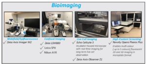
Bioimaging:
We have a range of microscopy platforms that facilitate the pursuit of fundamental biological questions pertaining to protein expression, localisation, and protein-protein interactions. We also have systems available for live cell imaging, and for high-content screening to evaluate morphological or viability changes in response to drug treatment or other modifying conditions.
The systems in Figure 1 are available in user-operated mode or, if assistance is required, staff-operated mode.
Figure 1
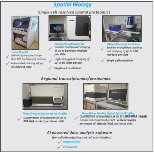
Spatial biology:
Our spatial biology capabilities enable the deep interrogation of both protein and RNA within formalin-fixed paraffin-embedded (FFPE) or fresh/frozen tissue, priming the discovery of novel cellular spatial relationships within the tissue, as well as the discovery/validation of clinically relevant biomarkers.
We provide fee-for-service options for the capabilities listed in Figure 2. User-operated mode is available for select units, please enquire at csi_spatial@nus.edu.sg for more details.
Figure 2
For further information, please email “Spatial Biology Core” csi_spatial[at]nus.edu.sg

Facility Head
Patrick William JAYNES
Core Facility Manager
pjaynes[at]nus.edu.sg
Team
Spatial Biology
Ms. Peng Yanfen
Senior Laboratory Executive
csipy[at]nus.edu.sg
mRNA/protein multiplex assays
Quantitative imaging and AI-based analysis
Ms. Tang Jing Ping
Senior Laboratory Executive
csitang[at]nus.edu.sg
Spatial transcriptomics/proteomics

Mr. Travis Lum
Research Assistant
trav.lum[at]nus.edu.sg
Multiplexed immunohistochemistry and spatial-omics assays
Bioimaging
Mr. Dedy Sandikin
Senior Laboratory Executive
dedys[at]nus.edu.sg
Advanced Bioimaging trainer & consultant
High content imaging and software analysis
Consultant

Dr. Anand D. Jeyasekharan
CSI Singapore
csiadj[at]nus.edu.sg